Foot drop (peroneal nerve palsy) - causes, symptoms & therapies

What is weak foot dorsiflexion?

The medical term “foot drop” is also known as foot drop palsy, peroneal palsy, peroneal nerve palsy, foot drop disease or drop foot. All these terms describe a condition in which the affected person has difficulty lifting the foot. This situation leads to the characteristic gait pattern known as “stepper gait”. The knee is raised unusually high to prevent the toes from touching the ground when walking – every step requires the utmost attention and there is a high risk of tripping or falling.

Foot drop can be caused by damage to the central nervous system (1st motor neuron: in the brain or spinal cord) or the peripheral nervous system (2nd motor neuron: from the spinal cord to the muscles).

If the cause is peripheral, the term peroneal nerve palsy or peroneal nerve paralysis is often used. This describes damage to the peroneal nerve (peroneus nerve), which is also known as the fibular nerve. Peripheral damage results in flaccid paralysis, which is an isolated paresis of the forefoot lifter muscles. In contrast, central damage causes spastic paresis, in which spasticity of the calf and paresis of the forefoot raiser muscles occur in combination.

Fußheberschwäche Fallfuß erklärt in Schema mit Peroneusnerv

Foot drop occurs when the strength of the muscles and nerves needed to lift the foot is lost. This weakness results from damage to the nerve and can occur in the brain, spinal cord or leg. In the case of foot drop (also known as peroneal palsy/peroneal nerve palsy), the brain can no longer correctly transmit the nerve impulses that control the lifting of the foot to the foot lifting muscles. The causes can vary: from a stroke, slipped disc or traumatic brain injury to a failed hip operation. Nerve diseases such as multiple sclerosis or Parkinson’s disease can also lead to weak foot dorsiflexion. In addition, the fibular nerve is susceptible to impact or crushing due to its course on the outside of the knee joint.

Foot drop manifests as a symptom due to nerve damage and describes the limited ability to lift the foot due to reduced strength in one or more of the following muscles:

  • Shin muscle (M. tibialis anterior)
  • Fibula muscle (fibularis longus/brevis muscle)
  • Toe lifter (extensor digitorum muscle)
  • Big toe lifter (extensor hallucis muscle)

The impairment of weak foot dorsiflexion is particularly noticeable during foot rolling and swinging the leg forward when walking. The forefoot, toes and/or lateral edge of the foot do not lift sufficiently or at all against the force of gravity(drop foot). This leads to a change in the gait pattern. The leg/knee must be lifted higher to prevent the affected person’s foot from sticking to the ground(stepping gait). Other adjustments to the gait pattern may also occur to prevent falls due to the foot catching. The persistent gait disorder leads to poor posture and pain over time. Early initiation of treatment is therefore recommended.

Terms at a glance: Foot drop, peroneal nerve palsy & co.

In short: foot drop mainly describes the symptom (the foot can no longer be actively lifted). Peroneal nerve palsy often refers to the cause (damage/paralysis of the peroneal nerve). Other terms are sometimes synonyms, sometimes accompanying phenomena in the gait pattern.

Foot drop (symptom)

Foot drop is the noticeable problem: the foot hangs in the swing phase – resulting in the typical drop foot. → More about treatment on this page.

Peroneal nerve palsy (cause, nerve lesion)

Peroneal nerve palsy is damage to the peroneal nerve, which often leads to weakness of the foot. Numbness on the back of the foot and a stepping gait are also typical.

Peroneal nerve palsy (synonym)

In practice, peroneal nerve palsy is usually referred to as the same clinical picture as peroneal nerve palsy – the focus is on the paralysis of the nerve.

Foot drop palsy (synonym with symptom focus)

Foot drop palsy emphasizes the weakness/paralysis of the foot lifting muscles. It is often the result of peroneal nerve palsy, but can also have other causes (e.g. L5 radiculopathy).

Drop foot & stepper gait (clinical picture)

Drop foot describes the dropping of the toe, stepper gait the compensatory lifting of the knee/hip when walking. These are observations, not the cause.

Symptoms & complaints of weak foot dorsiflexion

Walking on two legs is a natural but highly complex process: muscles/muscle groups in the foot, lower leg, thigh, hip and back have to interact with numerous joints as well as the senses of balance, sight and touch. In view of this, it quickly becomes clear how smoothly brain, nerve and muscle structures have to interlock here.

Orthopädie erklärt Fußheberschwäche am anatomischen Modell

In the case of weak foot dorsiflexion, damage to brain or nerve cells means that the nerve can no longer receive signals or transmit them to the muscle as a motor command. As a result, patients with weak foot dorsiflexion are unable to lift (dorsiflexion) and roll the foot when walking. A common symptom is the drooping toe of the foot (plantar flexion), which drags on the ground.

To compensate for the lack of mobility in the leg, a so-called stepper gait can occur: Those affected swing the impaired leg forward with a gyroscopic hip movement (circumduction) and lift it in the process. This unnatural gait can lead to pain and strain in the hips and spine, and can even lead to a slipped disc. Another consequence of the cramped posture is that patients often walk on the outer edge of the foot or claw with their toes. Walking is therefore no longer a natural process, but requires a great deal of attention with every step, and the risk of tripping and falling is extremely high.

Causes of weak foot dorsiflexion (neurological, muscular, traumatic, post-operative)

Foot drop can have various causes, such as damage to brain and nerve cells caused by a stroke, slipped disc or trauma. However, nerve diseases such as multiple sclerosis or Parkinson’s disease can also lead to foot drop – the brain can no longer control the lifting of the foot properly.

Below you will find an overview of various complaints and clinical pictures that can lead to weak foot dorsiflexion:

  1. Multiple sclerosis and weak foot dorsiflexion
  2. Foot drop after a stroke
  3. Foot drop in Parkinson’s disease
  4. Foot drop after hip surgery
  5. Foot drop after a herniated disc
Fußheberschwäche Ursachen Steppergang

Diagnosis of weak foot dorsiflexion (clinical, MRC scale, EMG/NLG, imaging)

Arzt stabilisiert Unterschenkel und prüft die Dorsalflexion des rechten Fußes (MRC) zur klinischen Abklärung einer Fußheberschwäche.

Clinical examination

  • Medical history: onset (acute/chronic), trauma/surgery, back pain, neuropathy risks.

  • Inspection & gait pattern: typical stepping gait, dragging toe, toe/heel gait.

  • Neurological status: Sensitivity dorsum of foot/1st interdigital space, peroneal pressure pain/tinula at the fibular head, reflexes.

  • Strength test (foot lift): Dorsiflexion (tibialis anterior muscle), big toe lift, eversion.

Degree of foot drop according to the MRC scale (strength grade 0-5)

  • 0: no contraction

  • 1: visible/palpable contraction without movement

  • 2: Movement only when gravity is switched off

  • 3: Movement against gravity

  • 4: Movement against low-strength resistance

  • 5: Normal force
    → Documentation of the foot lift (TA/EHL) on both sides, follow-up via re-tests.

Electrodiagnostics (EMG/NLG)

  • NLG peroneal nerve: distal latency, amplitude, conduction velocity; lateral comparison; detection of conduction block at the fibular head vs. axonal damage.

  • Sensitive (e.g. sural nerve): polyneuropathy indications.

  • EMG: Spontaneous activity (fibrillations/PSWs) from approx. 2-3 weeks, recruitment pattern, reinnervation potentials.

  • Differential diagnosis: L5 radiculopathy (EMG paraspinal/gluteal), plexus/ischiadicus lesion.

Imaging

  • Sonography peroneus: compression, ganglion, scars, subluxation at the fibular head.

  • MRI knee/lower leg: for trauma, suspected tumor/ganglion, surgery planning.

  • MRI lumbar spine: for radicular signs/therapy-resistant pain symptoms (L5/foraminal stenosis).

  • X-ray: suspected fracture/luxation.

Functional degree of foot drop in everyday life:

  • Easy: Inconspicuous gait, rarely stumbles

  • Means: Limited mobility, aids required

  • Severe: Permanent walking aid/orthosis, restriction of participation

Progression & prognosis of weak foot dorsiflexion

Progression of weak foot dorsiflexion

  • Acute (0-2 weeks): clinical stabilization, classification of the cause; with pure neuropraxia often rapid improvement.

  • Subacute (2-12 weeks): with axonal damage, denervation signs appear in the EMG (≈ 2-3 weeks), first reinnervation from ≈ 6-8 weeks.

  • Chronic (> 3 months): slow increase in strength through reinnervation; nerve regeneration approx. 1-3 mm/day. Fixed deficits → consider compensation strategies/surgery.

Prognostic factors

  • Favorable: mild paresis (MRC ≥ 3), short symptom/compression period, demyelinating conduction block without pronounced axonal loss, early increase in strength (≥ 1 MRC grade in 6-8 weeks), preserved/rapidly recurring CMAP amplitudes, younger age, no polyneuropathy.
  • Unfavorable: complete paresis (MRC 0-1) > 3 months, severely reduced/absent CMAP, pronounced denervation without recruitment after 8-12 weeks, persistent compression/trauma with axonotmesis/neurotmesis, diabetes/polyneuropathy, delayed therapy.

Control & Milestones

  • 4-6 weeks: clinical re-evaluation, EMG/NLG for localization & severity.

  • 3 months: no trend towards improvement → check cause, consider decompression/neurolysis if necessary.

  • 6-9 months: discuss tendinous procedures (e.g. tibialis posterior transfer) if there is hardly any reinnervation.

  • At any time: FES/AFO, physiotherapy, contracture and fall prevention ensure mobility and improve function – regardless of the aetiology.

Etiology-specific (short)

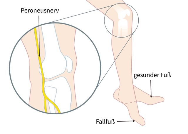
  • Peroneal compression/entrapment: often good recovery after pressure relief; months.

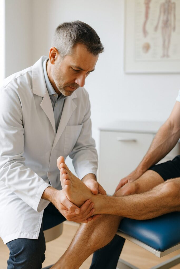
  • L5 radiculopathy: course depends on cause (prolapse vs. stenosis); clarify neurosurgically if deficit persists.

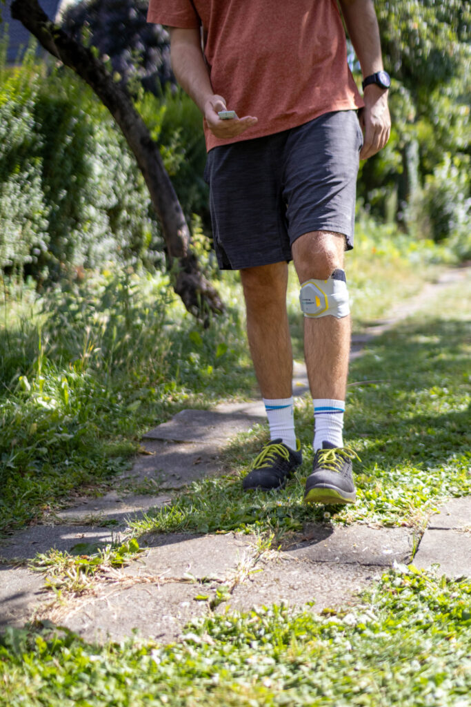
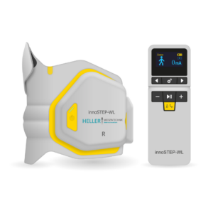
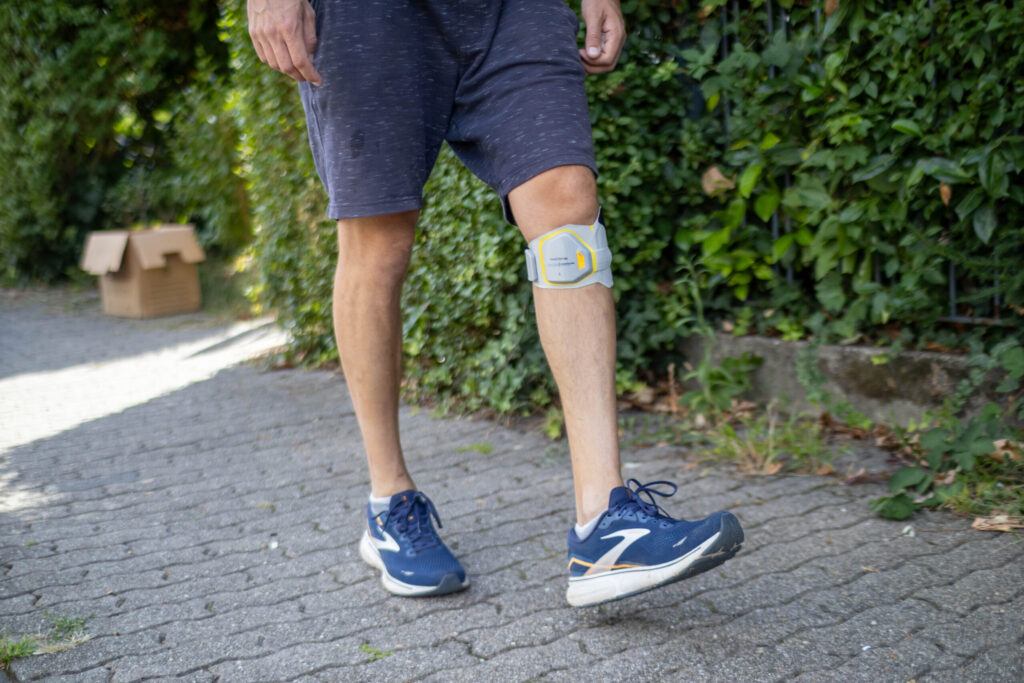
  • Central causes (e.g. stroke, MS): structurally persistent, functionally often partially compensated by neurorehab/FES.

Living with weak foot dorsiflexion

Depending on the cause of the paresis, there are different therapies and approaches.

It is essential to seek immediate medical help and a diagnosis if paralysis suddenly occurs. This is because life-threatening factors such as a stroke could be the cause. In such situations, the underlying disease must be treated first, which usually leads to relief of the symptoms of paralysis, i.e. paresis. Acute foot drop can also be caused by a herniated disc. In this case, it is just as important to seek medical assistance immediately. In many cases, prompt treatment/surgery is recommended in order to avoid lasting damage to the nerve.

As weak foot dorsiflexion usually occurs as a secondary injury, the medical focus is on treating the cause; the result can be an improvement in weak foot dorsiflexion. The innoSTEP-WL foot drop system, based on functional electrical stimulation (FES) , has proven itself as an aid for improving mobility and quality of life: Electrical impulses are sent to the peroneal nerve via an electrode cuff on the leg, which transmits them to the foot lifter muscle (tibialis anterior muscle) as a command to contract, causing the foot to lift during the swing phase (dorsiflexion).

The principle: electrical impulses are sent to the peroneal nerve (fibular nerve) via an inconspicuous electrode cuff on the leg. This transmits the signals to the foot lifting muscle (tibialis anterior muscle), which then actively lifts the foot during the swing phase(dorsiflexion). This creates a more natural, dynamic gait pattern that significantly reduces stumbling and falls.

Functional electrical stimulation offers further advantages over and above the immediate movement effect:

  • Promotes blood circulation, which supports healing and regeneration processes.

  • Prevention of muscle loss (atrophy) through regular activation of the shin muscle.

  • Training the nerve-muscle interaction: Repeated stimulus transmission and processing by the central nervous system promotes neuronal rewiring, so that functional improvement can occur in the long term even without stimulation.

This makes the innoSTEP-WL more than just a classic orthosis: it not only has a passive stabilizing effect, but also actively trains the muscles and nerve control. In the long term, this can even replace conventional peroneal splints or significantly reduce their use.


Comparison of treatment options for weak foot dorsiflexion

Physiotherapy & Training

Physiotherapy and training form the basis of the treatment: the aim is to increase strength, improve coordination and stretching ability and effectively prevent falls. This includes a structured gait analysis, functional exercises under everyday conditions and – where appropriate – supportive taping.

Foot lifter orthoses (peroneal splints)

Peroneal orthoses stabilize the foot immediately and are easy to use. However, their limitations lie in their mostly static mode of action: They prevent buckling, but only promote active muscle development and neuronal activation to a limited extent.

Functional electrical stimulation (FES) - innoSTEP-WL

Functional electrical stimulation (FES) with innoSTEP-WL starts earlier: A sensor detects the swing phase, electrical impulses activate the peroneal nerve or the foot lifters so that the toe lifts at the right moment. The result is a more dynamic foot lift, a more natural gait pattern and less stumbling; at the same time, the muscles and blood circulation are maintained and neuronal reorganization is promoted through repeated, correct movement. innoSTEP-WL is wireless, sensor- and remote-controlled, discreetly portable, suitable for everyday use and can also be used barefoot.

Surgical options for weak foot dorsiflexion

Surgical options such as tendon transfers or implanted stimulators are considered on an individual basis and are generally only considered after conservative measures have been exhausted.

innostepwl from Heller Medizintechnik in use for patients with weak foot dorsiflexion in the garden

Comparison of orthosis vs. FES vs. implant for weak foot dorsiflexion

CriterionOrthosisFES (innoSTEP-WL)Implant
Principle of actionPassively supportingActively stimulatingOperative, internal stimulation
Gait patternMore stable, rather rigidDynamic, more naturalDynamic
Muscle activityLowPromotedPromoted
CustomizabilityLimitedFinely adjustable, sensor-controlledDevice-specific
InvasivenessNoneNoneSurgical
Costs/reimbursementFrequentlyFrequentlyIndividual case
Criterion Orthosis FES (innoSTEP-WL) Implant
Principle of action Passively supporting Actively stimulating Operative, internal stimulation
Gait pattern More stable, rather rigid Dynamic, more natural Dynamic
Muscle activity Low Promoted Promoted
Customizability Limited Finely adjustable, sensor-controlled Device-specific
Invasiveness None None Surgical
Costs/reimbursement Frequently Frequently Individual case

Frequently asked questions about weak foot dorsiflexion

How can I recognize foot drop?

Those affected often stumble, have difficulty climbing stairs and typically have a dragging or stepping gait.

How is foot drop diagnosed?

The diagnosis includes a clinical examination, nerve measurements (EMG/NLG) and imaging procedures (e.g. MRI).

How can weak foot dorsiflexion be treated?

Foot drop can be treated with physiotherapy, special orthoses such as the innoSTEP-WL, electrical stimulation (FES) or – in severe cases – surgery.

What aids are available for everyday life with foot drop?

Aids such as foot lifter orthoses (AFOs), special shoes, electrical stimulation devices (FES) or trip protection in the home support those affected.

Can weak foot dorsiflexion be cured?

The chances of recovery depend heavily on the cause and duration of the weakness; early treatment significantly improves the prognosis.

How can I prevent weak foot dorsiflexion?

Good posture, regular exercise, targeted training of the leg muscles and the timely treatment of illnesses (e.g. diabetes, intervertebral disc problems) reduce the risk.

Who is FES suitable for?
  • Indications: Peroneal nerve palsy/falling foot after stroke, MS, incomplete lesions, peripheral nerve lesions, intervertebral disc pathology, etc.

  • Prerequisite: intact/responsive peripheral nerve pathway (EMG/NLG helpful), skin tolerance, realistic therapy goals

  • Contraindications (selection): implanted pacemaker/ICD (depending on the model), open skin lesions at the electrode site, unexplained tendency to seizures – clarify with a doctor

Is weak foot dorsiflexion the same as peroneal nerve palsy?

No. Foot drop is the symptom (the foot does not lift). Peroneal nerve palsy is a common cause (nerve damage). Many people use the terms synonymously – medically, the distinction helps with diagnosis and treatment.

innoSTEP-WL - Help with weak foot dorsiflexion for more freedom of movement

The innoSTEP-WL foot support system can be prescribed by your attending physician. You can send medical prescriptions directly to HELLER MEDIZINTECHNIK GmbH & Co. After approval by your health insurance company, you will be instructed in its use by a trained medical product consultant. You can also purchase the device.

We will of course be happy to advise you on fitting the innoSTEP-WL and other aids from the field of electromedicine: simply give us a call (+49(0)6442 9421-0), send us an e-mail or use our contact form. Further information and materials – such as a sample recipe – can be found here.

How do I get my innoSTEP-WL foot drop system?

The innoSTEP-WL is an aid for functional electrical stimulation for weak foot dorsiflexion, which can be prescribed by a doctor and covered by statutory health insurance. The necessary prescription can be issued by your family doctor or specialist. More and more doctors are giving you the option of requesting your prescription by phone or e-mail without having to visit the practice. Talk to your doctor. Depending on the patient’s insurance status, treatment is then provided in two different ways:

Legally insured persons

  1. The customer has a prescription issued via the innoSTEP-WL and sends it to HELLER MEDIZINTECHNIK GmbH & Co. KG (Europaplatz 2, 35619 Braunfels).
  2. HELLER MEDIZINTECHNIK prepares a cost estimate for a four-week trial and forwards it to the responsible health insurance company.
  3. After approval by the health insurance company, the insured person receives the innoSTEP-WL electrical cuff and is instructed in how to use it. Comprehensive patient documentation is created in accordance with the GKV guidelines, including a video of the gait pattern, with and without aids.
  4. If the insured person wishes to continue using the device after the four weeks have elapsed, they must send a follow-up prescription from their doctor to HELLER MEDIZINTECHNIK.
  5. HELLER MEDIZINTECHNIK submits a cost estimate to the respective health insurance company for permanent approval.

Privately insured persons

  1. The customer obtains a prescription for the innoSTEP-WL and orders the device from HELLER MEDIZINTECHNIK.
  2. HELLER MEDIZINTECHNIK prepares a cost estimate or an invoice for the purchase of the foot drop system (if required, also for a trial period of 4 weeks), which the insured person submits to his private health insurance company.
  3. Once the invoice has been settled, the insured person receives the innoSTEP-WL electrical cuff and is instructed in how to use it. Comprehensive patient documentation is created, including a video of the gait pattern with and without the aid.
  4. In the event of a trial, HELLER MEDIZINTECHNIK shall send the customer a cost estimate for the complete supply after the 4 weeks have elapsed.

Do you have any questions?

We will of course be happy to advise you on supplying the innoSTEP-WL: Simply call us on +49 (0)6442 9421-0, send us an e-mail or use our contact form. Further information and materials – such as a sample recipe – can be found here.

Cookie Consent with Real Cookie Banner